Many of you know that I am a major proponent and champion of 3D imaging in the general dentist practice. My original driving factor was purely dental implants. But more and more I am finding the most valuable use of 3D imaging is diagnosis and patient communication.
I’d like to share a patient that was just in my office for her routine hygiene recall visit. Amy has been in our practice for several years and due to various life issues has been taking care of her dental needs over time. This scenario isn’t uncommon in our practice – and likely most practices reading this.
In dentistry we have done a poor job by training our patients that dentistry is only needed when something is broken or hurting. In other words when there is a problem.
How many of your hygienists ask the question “Is there anything hurting or bothering you today?” at the beginning of their recall appointments. This single question drives me nuts and should be taken out of every hygienists vocabulary.
The most valuable tool I have found to overcome this is education using 3D imaging. The truth is that patients want to keep their mouths healthy, the issue is they don’t always trust us. To me nothing overcomes trust and skepticism more than visual proof – photographs and X-rays.
Here are Amy’s bitewing X-rays from her last visit 6 months earlier.


As you can see from the BWX, Amy has several things that need to be addressed. She has been putting these things off because they don’t hurt and they aren’t creating a pressing issue.
Here is Amy’s Panorex from the current visit.

Again, the panorex confirms the need to address some issues and take a closer look at a few other issues. What the panorex doesn’t show clearly is something that is happening in the right maxillary sinus. Take a closer look again at the panorex – do you see anything in the sinus. If you are like me you can’t really tell.
The great news here is that this ‘panorex’ is actually a 3D CBCT. This allows our office to dive deeper and take a much more detailed look at everything. In our office we have trained our hygienists to actually do this as well.
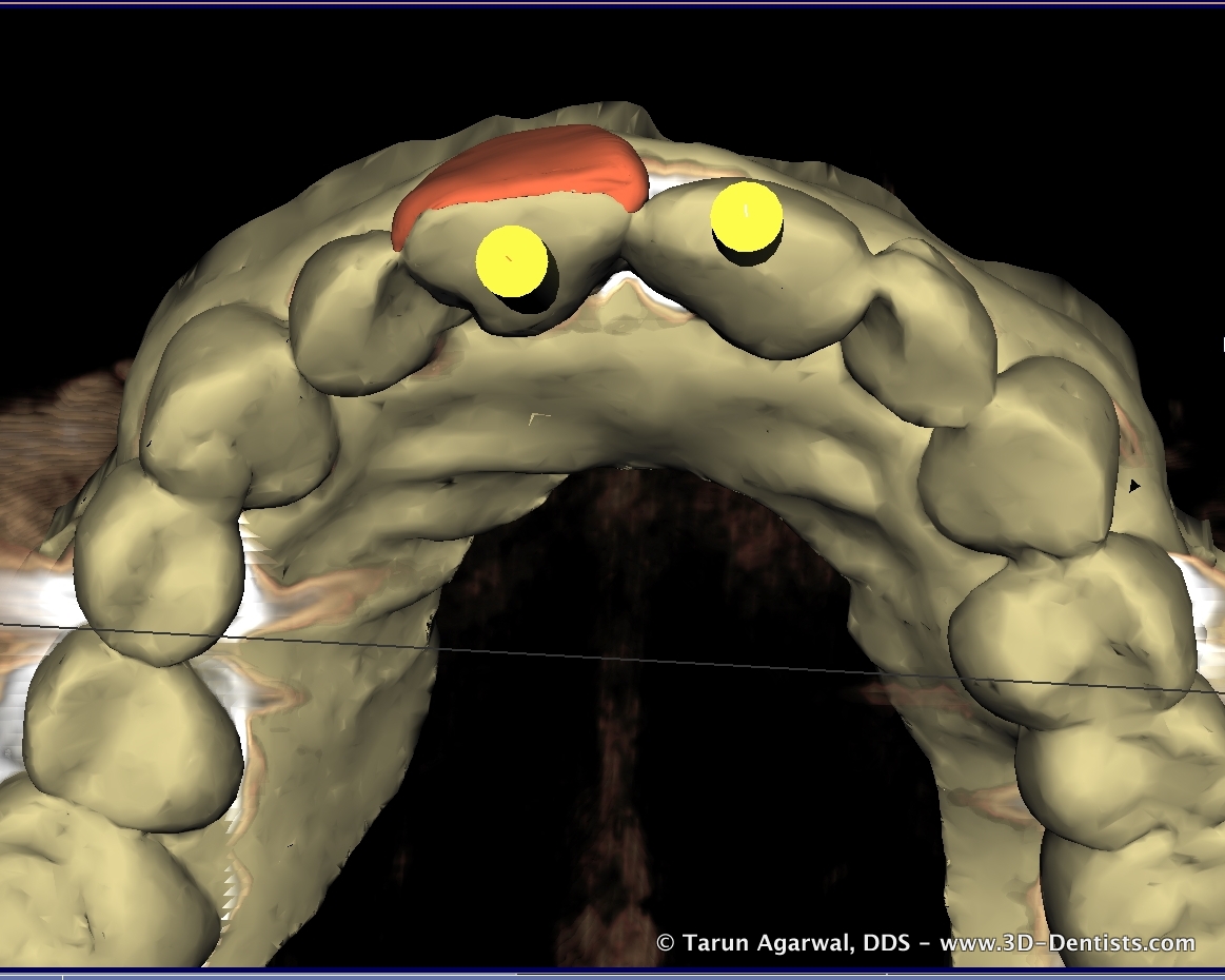
Here is screenshot of what my hygienist saw.

This image clearly shows there is something happening in the patients right maxillary sinus. She determines this not because she is trained to ‘read’ these images, but uses the concept of symmetry. The left sinus is totally clear.
When we see something in the sinus our first thought is to ask some questions. “Amy have you been experiencing some sinus issues recently? Specifically on your right side. The reason we ask is (and our hygienist points to the 3D image on a 40” patient monitor) right here we can see a big difference between your left and right sinus.”
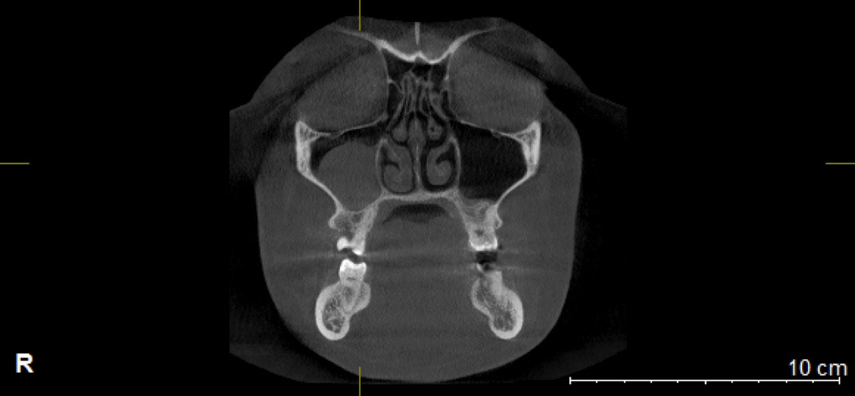
Our next step is to determine if the sinus issue it tooth in origin or if the patient needs to see their medical doctor. Here is a front screen shot of the area in concern.

Here what we see is the sinus fluid is just above the teeth and associated with the lower border of the sinus cavity. This leads us to take a closer look at the posterior maxillary teeth.
Here is a screenshot of a side view of the upper right teeth and the sinus.

Now we start to see that tooth #2 is a possible culprit. So now we take a much closer look. Here is what we find.

Now we clearly see that there is a peri apical lesion that has broken into the sinus. We next perform a ‘cold test’ on tooth #2 and find it to be non-responsive. This confirms the need for endodontic treatment on tooth #2.
Sooooo… who knows how long this has been going on. When we commenced treatment we found the tooth to be totally necrotic. It wasn’t hurting her because it was necrotic and had the ability to drain into the sinus.
This makes me wonder (and should make you wonder as well) how much we are missing because ‘it doesn’t hurt’. Fortunately/Unfortunately (depending on how you look at it), this type of diagnosis is now a normal for our office.
To help illustrate this case better I have created a video walkthrough of the 3D image. This allows you to dive in deeper with me and better understand the detail to which you can better diagnose and communicate with the patient.
Are you interested in learning more about this wonderful technology and how your patients and practice can benefit? May I suggest you attend a 3D Summit from Sirona Dental and Patterson Dental. These events are well done and a great experience. They bring in several fellow clinicians to share their stories and how 3D has positively impacted their practices. To learn more visit http://www.3DSummit.com . I am a featured speaker at every 3D Summit and would love to have you come by and say hello.